
The World-Leading Medical Imaging Platform for macOS.
OsiriX DICOM Viewer is Designed for radiologists, researchers, and imaging specialists, this software leverages the power of Apple’s hardware to provide the most advanced 3D and 4D rendering capabilities in the industry.
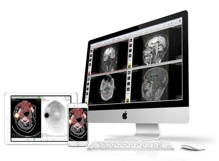
Unmatched Performance for Diagnostic Excellence
In the high-stakes environment of modern radiology, the ability to visualize and manipulate complex datasets quickly and accurately is paramount. The OsiriX DICOM Viewer sets the gold standard by fully utilizing the multi-core processors and graphic acceleration found in Mac computers. Unlike generic imaging solutions that struggle with large datasets, this application is built to handle thousands of images seamlessly, ensuring that patient diagnosis is never delayed by software lag.
At its core, the platform supports the complete DICOM standard for receiving, storing, viewing, and exporting medical images. Whether you are dealing with X-rays, CT scans, MRIs, or ultrasounds, the software provides a uniform, intuitive interface. The integration of advanced algorithms allows for immediate post-processing, meaning radiologists can adjust window levels, apply filters, and generate 3D reconstructions instantly. This level of responsiveness is critical when time is of the essence, such as in emergency trauma centers or during complex surgical planning sessions.
Beyond mere visualization, the application excels in multimodality fusion. Users can overlay PET (Positron Emission Tomography) images with CT or MRI scans to create a comprehensive view of metabolic activity combined with anatomical structure. This fusion capability is indispensable in oncology for pinpointing tumor locations and assessing treatment response. The platform also supports 4D rendering for dynamic sequences, such as cardiac function studies, allowing clinicians to observe the beating heart or flowing blood vessels in real-time.
Advanced Visualization Tools
A complete suite of tools designed for the modern radiologist.
3D & 4D Rendering
Generate high-resolution 3D volume renderings from CT or MRI series. Visualize complex anatomical structures with depth, shading, and perspective.
PET-CT Fusion
Overlay functional and anatomical images. Automatically register and align datasets to enhance diagnostic accuracy in oncology and neurology.
Heuristics & AI
Utilize advanced lesion detection algorithms. The software automatically identifies potential regions of interest to assist radiologists during the reading process.
Complete DICOM Support
Read, display, and analyze all DICOM file types. Support for compressed images, JPEG 2000, and various proprietary formats from major manufacturers.
PACS Integration
Query and Retrieve (Q/R) from any DICOM server. Seamlessly connect to hospital infrastructure to download studies directly into your local database.
Extensible Plugins
Expand functionality with a robust plugin architecture. Add custom algorithms, support for research protocols, or specialized measurement tools.
Optimized for macOS Architecture
Many cross-platform imaging solutions fail to take full advantage of the hardware they run on, resulting in sluggish performance and poor image quality. In contrast, the OsiriX MD software is natively developed for macOS. This native foundation allows it to utilize Metal, Apple’s high-performance graphics framework, to deliver smooth 60fps navigation even through voluminous datasets.
The software supports 64-bit computing, allowing it to address massive amounts of RAM. This is essential when stacking hundreds of CT slices or performing complex deconvolution algorithms on MRI sequences. Furthermore, the application is fully optimized for the latest Apple Silicon (M1/M2/M3 chips), delivering unprecedented energy efficiency and battery life for mobile radiologists reading on MacBook Pros or iPads.
✓ Multi-touch gesture support for intuitive navigation
✓ Full-screen mode for distraction-free reading
✓ Direct support for Retina and ProMotion displays
Frequently Asked Questions
OsiriX DICOM Viewer is an advanced medical imaging software designed for viewing, processing, and analyzing DICOM images from CT, MRI, PET, Ultrasound, and other imaging modalities. It enables healthcare professionals to perform efficient image interpretation, 2D/3D visualization, and diagnostic analysis.
Yes, the MD version of the OsiriX DICOM Viewer has received 510(k) clearance from the U.S. Food and Drug Administration (FDA) for medical image diagnostic use. This ensures that it meets rigorous standards for safety and effectiveness required in clinical environments.
Absolutely. The software includes a dedicated module for creating standardized DICOM media. You can easily select a patient study and burn it to a disc, complete with a built-in cross-platform viewer (compatible with Windows) to ensure referring physicians can open the files immediately.
There are two main versions available: “Lite” is available for free and is intended for educational purposes, image viewing, and basic research, though it cannot be used for primary diagnosis. “MD” is the licensed, paid version that includes all advanced features, FDA clearance, and technical support.
OsiriX helps healthcare professionals quickly access, visualize, and analyze medical images, enabling faster diagnoses, improved collaboration, better treatment planning, and enhanced patient outcomes through efficient and accurate medical imaging workflows.
See Beyond the Image
Empower radiologists, clinicians, and healthcare professionals with advanced DICOM visualization technology designed for faster image interpretation, diagnostic confidence, and improved patient care.